
Pregnancy is an exciting time for most women. As the pregnancy progresses, you may wonder about your baby’s development.
You cannot wait to tell the good news to your family and schedule an ultrasound to show a sonogram image of the baby.
An early ultrasound can be necessary; a lot of development happens with the baby after 7 weeks. You may feel a mix of emotions at the result of your ultrasound.
There are growth factors in the baby you can see to detect signs of life. However, the features of the baby will not be evident yet.
An early pregnancy dating scan will help assess the expected date of delivery. It can also rule out pregnancy complications and help you evaluate your health.
A 7-week ultrasound will most likely be your first.
It would help to know what to expect and what the sonogram image will show you. You will have queries about your pregnancy.
If you have an idea of what to expect in your ultrasound, it will help you know what questions to ask your doctor.
What to Expect at Your 7 Weeks Pregnancy Ultrasound
What is an Ultrasound Scan?
Ultrasound in pregnancy is the standard method of monitoring the health of you and your baby.
The use of ultrasound to evaluate the status of a woman’s pregnancy has increased in high numbers from 1995 to recent years.
The purpose of an ultrasound is to monitor the status of your pregnancy and diagnose acute trauma or complications.
Ultrasounds have been portrayed in visual media such as television, movies, social media, etc.
It is the most common diagnostic procedure in obstetrics. It is a safe, convenient and painless process that yields immediate results.
The purpose of ultrasound during routine prenatal care is to confirm the baby’s presence. The gestational age, heartbeat, and number of fetuses can be determined at 7 weeks.
Your obstetrician makes a thorough diagnosis to ensure the safety of your pregnancy. Ultrasounds, blood tests, and other examinations are performed to identify the causes of suspected complications.
An ultrasound can be done through two different methods:
Transabdominal ultrasound scan
It is an external examination where an imaging device called a transducer is placed on the lower abdomen to create a sonogram image of the uterus’s interior.
This procedure is done by placing a cool gel across the pregnant woman’s belly and using a transducer device to generate a picture of the baby developing in the womb.
Sound waves are utilized to project an image on a screen similar to a live feed for viewing and recording.
Nevertheless, transabdominal ultrasound is more suited for later on during the pregnancy than early since the baby would generally be bigger and easier to examine through this method.
Transvaginal ultrasound scan
It is an internal examination.
In this method, the transducer is placed into the vagina. Transvaginal scanning is meant for earlier stages of the pregnancy when the embryo is still very small.
This method calls for inserting the transducer inside the vagina. In doing so, this method can produce better quality images of your growing baby than with a transabdominal ultrasound.
Why do I need an Ultrasound Scan at 7 weeks?
Healthcare providers would typically request that you take an ultrasound for more accurate dating of your pregnancy, especially if you don’t know the exact date of conception.
For even more accurate measurements of an embryo, it is recommended that you have an ultrasound done during your first trimester rather than later on in your pregnancy.
A sonographer can use the measurements taken to pinpoint a baby’s age.
This is because the varying growth rates of a baby make it less reliable to predict a baby’s age.
Using these measurements to determine an embryo’s age would be less reliable the larger the embryo develops in the womb. Similar to how children vary in shape and size.
Your health care provider might ask you to get an ultrasound at 7 weeks to:
- Confirm the presence of the baby
- Check if you have a twin or multiple pregnancy, especially if you have had fertility treatments.
- Check the baby’s heartbeat
- Check for ectopic pregnancy
- Check for complications if you’ve been experiencing problems such as pelvic pain and vaginal bleeding, etc.
- Measure the size of the embryo
- Assess the embryo’s growth if there is uncertainty about the date of your last normal menstrual period.
- As a general check-up on the health of the mother’s uterus, fallopian tube, ovaries, and pelvic organs.
A 7 weeks pregnant woman can experience various symptoms. Some common and harmless symptoms include nausea, morning sickness, and cravings.
Despite this, if you’ve had concerning symptoms associated with your pregnancy, such as spotting or bleeding, your doctor will ask for an ultrasound.
Some unpleasant symptoms during pregnancy may be signs of miscarriage or ectopic pregnancy.
Issues with your uterus, cervix, ovaries, or fallopian tubes can complicate your pregnancy. Your doctor will do a general check-up of your reproductive system to ensure that you and the fetus are healthy.
An early ultrasound will help you be reassured of your reproductive health and the growing embryo.
Mothers can benefit from accurate dating of their pregnancies, as your doctor would be able to make better decisions about how to carry on with your pregnancy.
This includes women going into labor earlier than the normal 9 months (premature birth) or a little later after 9 months (overdue birth).
Doctors can determine accurate dating because an ultrasound will give them a good early peek at the mother’s developing baby as it grows.
You will have to inform your doctor about your medical history, existing conditions, symptoms, and medicines. This information includes if you recently stopped using oral contraceptive pills.
What to Expect at a 7-week Ultrasound
A 7-week ultrasound is usually recommended by a medical professional to collect information on your health.
During the examination, two types of equipment can be used for an ultrasound—a transvaginal probe for a transvaginal scan and a curvilinear probe for a transabdominal scan.
Before the ultrasound, you will need to have a full bladder for a transabdominal scan at 7 weeks.
A transvaginal ultrasound will not require a full bladder, but you will need to be ready for the insertion of the probe into the vagina.
A pregnancy dating is from the first day of your last menstruation, and the average duration of pregnancy is 280 days.
Pregnant women find the first signs of life in a baby in the early weeks of the pregnancy.
The sonographer will scan for the gestational sac with the embryo to detect if the location is within the uterus. A heartbeat will then be found to confirm signs of life.
The number of gestational sacs and embryos in each sac are counted to assess how many babies are present.
A single pregnancy will have one gestational sac, and multiple pregnancies will have two gestational sacs with an embryo each.
However, having more than one baby may also include one gestational sac with more than one embryo within.
The pelvic region will be scanned to ensure that you can conceive without complications. Furthermore, the gestational age of the embryo is determined from crown to rump measurement.
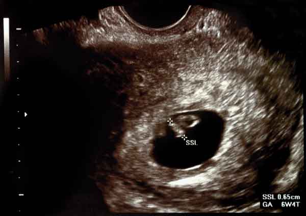
What a 7-week Pregnancy Ultrasound Would Look Like
In the first weeks of pregnancy, you will barely see a resemblance of your baby’s features.
Nevertheless, some indicators are essential to evaluate the status of your baby. A duration of one week can make a significant difference in the development of the embryo.
During the week 7 ultrasound, your baby is about the size of a blueberry at a few millimeters.
At this time, you’ll be able to figure out your due date and have an ultrasound to check for the baby’s heartbeat.
Fetal viability is confirmed once the sonogram image shows an embryo with cardiac activity. A heartbeat is usually detectable at the 6th week of gestation when the embryo is 2 mm or greater.
Other sonographic features of early pregnancy can also predict the baby’s presence. The first evidence of pregnancy found in ultrasound is the gestational sac.
At this stage in the pregnancy, the baby’s heartbeat steadily increases, beating faster than it did the day before.
And as your baby takes form from a tiny spec at 7 weeks, they would now have their limbs developing in the form of little flippers with joints at the 8th week.
In the following weeks, facial features such as eyes, ears, tiny hands, toes, and nostrils would also start to show.
Measurements Taken of the Baby’s Growth
If you can’t pinpoint the exact date of conception, the best way to chart your pregnancy is by counting forward from when you last had your last menstrual period, assuming that you’ve ovulated 14 days after your period began.
Doctors would benefit from this information and measurements, for it would be important for the timing and interpolation for specialized prenatal tests.
Quadruple screen, nuchal translucency measurements, and others are used to assess whether or not genetic abnormalities are developing.
1) Gestational Sac
The sonograph image will show the gestational sac at the center of the uterine body at 4.5 to 5 weeks as evidence of the embryo’s presence. It is the first structure visualized in pregnancy.
The gestational sac is the fluid-filled round structure surrounding an embryo during the first weeks gestation of the baby.
When a woman is 7 weeks pregnant, the gestational sac is found in the central echogenic portion of the uterus.
It will be at 2 to 3 mm in diameter and increase by 1.13 mm per day reaching 7-8 mm at 7 weeks.
2) Embryo
At 5.5 weeks, a yolk sac would be found within the gestational sac. The yolk sac is a thin-walled circular structure representing the embryo with the gestational sac.
Embryo size is measured using crown-rump length assessment.
This is the most accurate method of measuring the gestational age of the embryo in early pregnancy. In the 8th week of gestation, the head, body, and limb buds will start to grow.
Unfortunately, these will not become apparent in the ultrasound of 7 weeks pregnant women.
3) Cardiac Activity
The fetal heartbeat is a good indicator of the baby’s health. It is seen as a flickering structure in ultrasound at 7 weeks.
Additionally, a strong fetal heart rate reassures that your baby is healthy.
Six or seven weeks are the earliest time to detect cardiac activity. This will show up as a pulsing motion within the embryo.
Complications in Pregnancy
A gestational sac is usually the first thing you’ll see in a healthy pregnancy.
However, there are circumstances that you may not see the yolk sac, fetal pole, shape of your baby, or the heartbeat yet.
This may only mean that you are still early in your pregnancy than you predict. Additionally, a tilted uterus can be an issue in performing an ultrasound.
It can make it harder to see the baby until they’re a little bigger.
The position of your baby can affect how the measurements are taken.
Even the number of gestational sacs and embryos can be inaccurate when one embryo can hide behind another.
A 7-week ultrasound becomes crucial when possible complications can be pointed out. The earliest weeks of pregnancy have the highest risk of miscarriage.
Inconsistent signs of pregnancy can show up, such as a large gestational sac without any yolk sac or fetal pole.
Nevertheless, your worries may be caused by an inaccurate dating of your pregnancy.
1) Ectopic pregnancy
It is when an embryo grows outside the uterus. You will still experience early pregnancy symptoms and have a positive pregnancy test.
It is a life-threatening condition for pregnant women and their babies.
An ultrasound is performed when concerning symptoms such as vaginal bleeding, abdominal pain, or pelvic pain are found in a 7-week pregnant woman.
This is to rule out the possibility of an ectopic pregnancy.
Your health care provider may ask for a blood test and pelvic exam if there is no fetal growth in your uterus.
Other factors assessed include intrauterine pregnancy, pregnancy of unknown location, pregnancy viability, and the fetal heartbeat.
2) Transvaginal Ultrasound Contraindications
It may be contraindicated in high-risk patients.
Special considerations are made in patients with acute vaginal bleeding and unknown past medical history.
In these cases, a transabdominal scan is performed first. Once a doctor deems it safe, a transvaginal scan is completed.
Generally, it is safe to execute even in the later weeks.
The angle between the cervix and the vaginal probe prevents it from slipping into the cervix and disrupting the placenta.
The Bottom Line
The early weeks of pregnancy may not seem as crucial to the growing baby.
However, the seven weeks of the first trimester hold essential indicators for a mother and the baby.
The measurements found in an ultrasound of a 7 weeks pregnant woman will accurately predict the estimated due date compared to later weeks.
Ultrasound is routinely performed during the development of babies.
It can make a big difference to monitor your baby’s growth and how it’s taking shape in your body.
The baby may show as a speck in the sonogram scan, but the first sign of life will appear in the first trimester.
You will see the embryo’s size and the baby’s heart beating at 7 weeks.
Although getting an ultrasound as early as the 7th week is beneficial for both the mother and the doctor, it isn’t 100% guaranteed that doctors can catch problems on the first ultrasound.
It is usually because the baby is still growing.
Subsequently, a woman will have 2 more ultrasounds throughout the pregnancy.
Another ultrasound will be done on the 11th-13th week or the second trimester as a general examination of the baby.
A follow-up on the 18th-20th week or the third trimester is done to determine the gender of the child.